Support in Insurance Claim
No-Cost EMI
Without Admission
Short Hospital Stay
4.9 Rating on Google
Benefits

Minimally Invasive

No Complications

Faster Recovery

Protects Brain Function
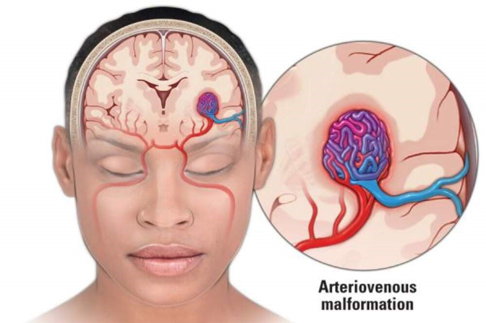
What is a Brain AVM?
A brain Arteriovenous Malformation (AVM) is an uncommon abnormal cerebral blood vessels. It interferes with normal blood flow through direct connections between veins and arteries that bypass the vital capillary system. This condition causes high-pressure blood flow in weak vessels, which are more prone to rupture. If untreated, the AVMs in the brain pose serious risk to health as the weak vessels can rupture in a way that could cause bleeding within the brain (hemorrhage) or stroke or irreparable neurological damage. Early diagnosis and treatment in a specialist AVM hospital such as IRFacilities are vital to prevent these serious problems and maintaining the neurological function.

Understanding Brain AVM Symptoms
A cerebrovascular arteriovenous malformation (AVM) is a rare blood vessel knot in the brain. The symptoms it creates depend on the dimensions and exact location.
Some people do not show any symptoms and are unable to tell if they have an AVM exists. In some, it could result in noticeable neurological issues.
Common Symptoms to Be Aware Of:
- Headaches: Headaches are typically intense persistent and can be different from typical headaches.
- Seizures: They can be triggered by sudden periods of convulsions or staring spells, or bizarre sensory sensations.
- Numbness or Weakness: A sudden or gradual loss of feeling or strength usually in one leg or arm.
- Speech Issues: Difficulty forming words and understanding what people say, and identifying the correct words.
- Vision changes: It could be an impairment of your vision blurred vision, seeing double.
Causes and Risk Factors
The exact reason behind the brain's arteriovenous malformations (AVMs) is not entirely known. They are typically regarded as congenital which means they occur as a result of mistakes in the formation of blood vessels. Although not often passed down through the family, a genetic link can be found in certain conditions.
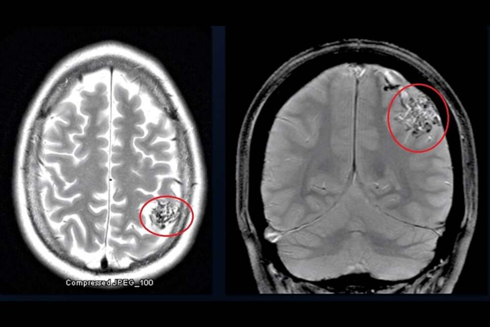
There is no evidence of cause of this condition that is not a result of environmental or lifestyle factors. Thus, the diagnosis is crucial and heavily depends on the area that is AVM Radiology. Modern imaging techniques such as MRI as well as CT angiography are employed to precisely determine the size of the malformation and location as well as pattern of flow. The precise radiological assessment is vital for precise guidance in subsequent treatment plans and to ensure a safe treatment strategy.
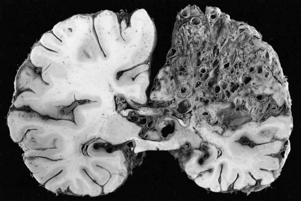
Brain AVM Types and Urgent Red Flags
The brain AVMs can be classified by their place of origin and blood supply, which determines the treatment plan. The most common kinds are:
- Pial AVM is situated on the top of the brain. It is supplied via branches of the internal carotid artery (ICA)
- Dural AVM is located inside the tough outer layer of the brain (the dura) and is supplied via some branches from the external carotid artery (ECA)
- Mixed AVM: Provided via branches of both the ICA and ECA
Sure signs, referred to by the term "red flags," indicate the possibility of rupture and need prompt or urgent treatment. They include:
- A prior history of AVM bleeding.
- A diagnosis of an intracranial aneurysm or an arteriovenous (AV) fistula in the AVM
- Atypical, small bulges that are located within the drainage veins (venous pouches)
- Uncontrolled seizures triggered by the AVM
Treatment Options for Brain AVM
We are at IRFacilities We focus on the most advanced, minimally-invasive treatment options to treat neuro arteriovenous malformations (AVMs). Our neurointerventional specialists are highly skilled and ensure that each patient receives treatment specific to their needs.
AVM Embolization
For the majority of patients, it is the best procedure. This procedure is safe and catheter-based. It is a better alternative to open surgery. A tiny tube can be inserted directly through the blood vessels into the AVM. There, specific agents are employed to stop blood flow that is not normal.
We use two proven techniques:
- The Glue-Based Embolization (NBCA): Our preferred method, in which medical-grade glue is used to seal off any abnormal vessels.
- Squid Embolization/Onyx: For cases that require the exact placement of liquid embolic material.
These techniques help to control bleeding, minimize risk, and, in certain instances, cure AVMs completely. AVM completely. They can also help make other treatments more secure when needed.
Other Treatment Options
In certain instances, alternative options may be considered
- Surgical removal is indicated for AVMs that are safe and accessible to perform surgery on.
- Stereotactic Radiosurgery is a non-invasive method for AVMs with small diameters and deep-seated locations, although recovery times are more sluggish than embolization.
Why IRFacilities?
We believe that embolization is the most effective combination of safety, effectiveness, and speed of recovery. Utilizing the most advanced techniques and skilled experts, IRFacilities provides patients with superior outcomes and quicker recovery times than conventional options.

Recovery Time and Success Rate
The recovery process following AVM embolization performed at IRFacilities is much more rapid than open surgery. Patients are typically required to remain at our AVM hospital to be monitored for 1 to 3 days after the procedure. The majority of patients can resume their routine activities in one or two weeks although strenuous activity may be limited for a brief time. Recovery is typically quick because of the non-invasive procedure, which requires a minor puncture on the groin.
The procedure boasts a chance of success with a rate of between 80 and 90 percent in effectively preventing the flow of blood to the tumor and prevents rupture of AVMs in the brain. If combined with radiosurgery or surgery, the rate of total AVM elimination is much more impressive. Utilizing advanced imaging and a small brain catheter enables exact delivery of embolic drugs that result in fewer complications, less pain and superior long-term outcomes for patients, confirming its status as a leading treatment option.
AVM Embolization Cost in India
The AVM embolization cost in India at IRFacilities is significantly more affordable than in Western countries, making us a leading destination for high-quality brain AVM care. The final expense depends on the technique and materials used:
- Glue-Based Embolization: ₹1.5 – 2 Lakh. Our most cost-effective solution is ideal for standard cases where medical-grade glue provides optimal results.
- Onyx/Squid Embolization: ₹3 – 4.5 Lakh. A premium option for complex brain AVMs, utilizing advanced materials that allow superior precision and control in challenging cases.
Despite these limitations, AVM embolization at IR facilities is still a fraction of the expense of traditional open-brain procedures. It offers world-class treatment without sacrificing the quality of care or results. The entire procedure from diagnosis to procedure itself is based on advanced AVM radiology to guarantee the safety and efficacy, as evidenced by the total effectiveness of the procedure.