

Support in Insurance Claim
No-Cost EMI
Without Admission
Short Hospital Stay
4.9 Rating on Google
To Book An Appoinment
Introduction of Prostate Artery Embolization (PAE)
Prostate Artery Embolization (PAE) is a relatively new minimally invasive therapy for men with benign prostatic hyperplasia (BPH), which is the non-cancerous enlargement of the prostate gland. As men age, particularly past the age of 50, enlargement becomes increasingly commonplace, and it may produce disagreeable sensations such as waking up at night to urinate, initial hesitation or interruption of urine flow, or a sensation of not fully emptying the bladder.
PAE is a non-invasive procedure that reduces the size of the prostate by diminishing its blood supply; it is performed by a specialist called an interventional radiologist. Through a tiny puncture in the wrist or groin, the doctor accesses the arteries supplying the prostate via a tiny catheter. Particles are injected through these arteries to block the blood supply. Over time, the prostate shrinks and, along with it, improvements of symptoms.
PAE has some advantages: no surgery, shorter recovery time, and less risk of possible side effects such as erectile dysfunction as compared to the traditional treatments. Most patients would go home within a day after undergoing the procedure, and return almost immediately to normal activities in about a few days.
It is a safe as well as effective procedure for men who are interested in relief of BPH symptoms using a non-invasive approach to improve their quality of life.
Prosthetic Artery Embolization at IRFacilities: A Step-by-Step Guide
At IRFacilities, prosthetic artery embolization (PAE) is a meticulously planned and executed procedure, ensuring patient safety and optimal outcomes. Here's a detailed overview of how the process unfolds:
Initial Evaluation on an Outpatient (OPD) Basis
- Comprehensive Counseling:
- During the OPD visit, the patient undergoes a thorough consultation. They are educated about the procedure, its benefits, and potential risks. This session also addresses any patient queries.
- Diagnostic Imaging and Planning:
- A urodynamic study and a CT-ANGIO of the pelvis are scheduled.
- A low-pitch CT scan with IMR (Iterative Model Reconstruction) at 0.6 mm resolution is conducted. This advanced imaging provides a highly detailed anatomy of the prostatic artery, which is often variable.
- The Bull’s Tracking Method is used to trace the vascular anatomy with precision.
- Pre-CT Assessment:
- Creatinine and baseline tests are evaluated to ensure kidney function can safely support contrast imaging.
- PSA (Prostate-Specific Antigen) levels and PSA velocity are measured to rule out any prostate malignancy.
- A trus transducer ultrasound is performed to cross-verify prostate size and anatomy.
Team Review and Anatomy Analysis
- Detailed Arterial Mapping:
- Whether the prosthetic artery is single or double on either side.
- The origin of the prostatic artery, which typically arises from the pudendal artery, obturator artery, or superior vesicular artery. Any aberrant origins are also noted.
- Collaborative Assessment:
- Junior IR specialists first map the anatomy, followed by a senior IR for confirmation, ensuring no anatomical detail is overlooked.
- Prostate Size Measurement:
- The prostate's size is assessed using both CT imaging and transrectal ultrasound (TRUS) for accuracy.
The imaging results are analyzed by the interventional radiology (IR) team to identify:
Pre-Procedure Evaluation
- Comprehensive Health Check:
- Any abnormalities, such as malignancies or other conditions apart from benign prostatic hyperplasia (BPH), are ruled out.
- Urodynamic Study and Uroflowmetry:
- This study is conducted to evaluate bladder function and any associated urological concerns.
- Patient Follow-Up:
- The patient is sent home and waits for a call back from us later for the actual embolization procedure.
Why Choose IRFacilities for PAE?
The expertise and precision of the IR team at IRFacilities ensure that every aspect of prosthetic artery embolization is carefully planned. From cutting-edge imaging techniques to collaborative assessments, the focus remains on delivering effective, minimally invasive treatment for BPH while prioritizing patient comfort and safety.
Prostatic Artery Embolization (PAE): Procedure Overview at IRFacilities
At IRFacilities, prostatic artery embolization (PAE) is performed with precision and patient comfort as top priorities. Here's what to expect during the procedure:
Pre-Procedural Preparation
- Arrival and Fasting:
- Patients are instructed to report on the morning of the procedure and remain fasting from 5 a.m. that day. To ensure a calm state of mind, a 0.5 mg dose of is advised.
- Safety Assessment:
- Elderly patients with comorbidities like cardiovascular or neurovascular conditions are evaluated thoroughly by the sedation team. Risks are minimized, and procedures are only carried out after obtaining informed consent.
- Pre-Op Area:
- The patient is provided an operating theater (OT) gown and attended by interventional radiologists (IR) and nursing staff.
- An intravenous cannula is inserted, usually in the left hand or arm, and fluids are started.
- The patient is counseled about the minimally invasive nature of PAE, which involves no significant pain.
- Groin Preparation:
- The groin is prepared for access unless religious reasons, such as those of the Sikh community, necessitate leaving the hair intact.
During the Procedure
- Cath Lab Setup:
- The patient is moved to the cath lab at the scheduled time. Non-invasive monitors are attached to track vital signs, and the patient’s blood pressure and overall stability are checked. The patient is placed in a comfortable supine position.
- Sterile Preparation:
- Both groins are painted and draped to maintain sterility.
- The right groin is typically used for access, with the left groin only being utilized in specific cases.
- Femoral Artery Access:
- A local anesthetic is applied liberally to ensure the patient’s comfort.
- A 6-French sheath is inserted into the right femoral artery, with the patient informed at every step to maintain confidence and comfort.
Prostatic Artery Embolization (PAE): Detailed Procedure at IRFacilities
Step-by-Step Procedure
- Accessing the Femoral Artery:
- A 6-French sheath is placed in the right femoral artery, which serves as the entry point for the procedure.
- Using a Terumo 0.035 guidewire, an RC2 catheter is advanced into the left common femoral artery.
- Navigating to the Prostatic Artery:
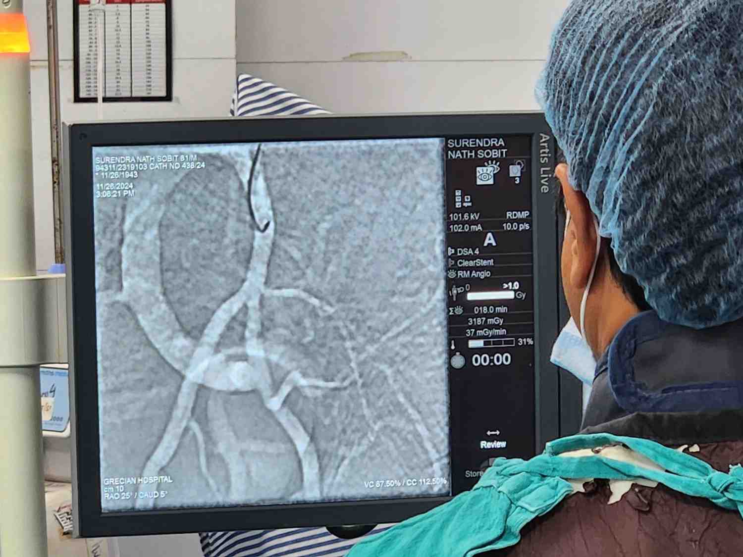
- The RC2 catheter is further guided into the internal division of the internal iliac artery based on information obtained from a pre-procedure CT angiogram (CTAngio).
- The CTANGIO helps locate the variable origin of the prostatic artery, minimizing the need for extensive exploration during the procedure.
- Once the origin of the prostatic artery is identified, the tortuosity of the artery is assessed, often using a contralateral oblique view for better visualization.
- Microcatheter Placement:
- The RC2 catheter is positioned slightly proximal to the prostatic artery's origin.
- A microcatheter, typically an 0.017-inch ID microcatheter paired with a 0.014 guidewire, is introduced. The tip of the microcatheter is steam-shaped, and the guidewire is needle-shaped for precise navigation.
- Using a "roadmap" image obtained before microcatheter advancement, the microcatheter is carefully maneuvered deep into the prostatic artery.
- Prostatic Artery Embolization:
- After advancing the microcatheter to the desired position, the guidewire is removed. Contrast is injected to confirm the microcatheter's position through parenchymal staining of the prostatic artery.
- Embospheres, typically 75-150 microns in size, are delivered through the microcatheter. These small particles penetrate deeply into the prostatic tissue, effectively reducing blood flow to the gland.
- The microcatheter is gradually withdrawn to embolize additional branches of the prostatic artery as needed.
- Treating the Contralateral Side:
- The procedure is repeated on the right side using either a Robertson Uterine Catheter (RUC / UAC) catheter from the same groin.
- If contralateral groin access is required, an RC2 catheter is again used, and the process mirrors the left-side embolization.
- Completion:
- After embolizing both prostatic arteries, all catheters are removed, and the femoral artery is closed.
- The patient is monitored post-procedure before being discharged, often on the same day.


Our Services
- Varicose Veins
- Varicocele
- Brain Hemorrhage Interventions
- Brain Hemorrhage
- Aneurysm coiling
- SAH vasospasm management
- Flow diverter placement
- AVM embolisation
- Pancreatitis
- Pancreatitis Interventions
- Trans visceral drainage
- Uterine fibroid embolisation
- Hemoptysis Interventions (BAE)
- Portal hypertension Interventions (TIPS ,BRTO)
- Ascites intervention
- Pleural effusion intervention
- Biopsy /FNAC
- Liver abscess drainage
- Bleeding Embolisation
- Splenomegaly Embolisation
- Biliary Drainage
- Stroke Interventions
- Liver Cancer
- Metastatic Cancer
- Pain Management
- Peripheral artery management
Make An Appointment
Post-Procedure Care for Prostate Artery Embolization (PAE):
The minimally invasive procedure of Prostate Artery Embolization (PAE) reduces the size of the prostate for symptom relief in benign prostatic hyperplasia. Optimal post-procedural care ensures better recovery results.
Immediately After the Procedure:
Following PAE, the patient will be under observation on an outpatient basis in the recovery area for several hours to provide adequate time for patient to start feeling normal.
- Pain Management:
- Hydration:
- Activity Restrictions:
After PAE, it is quite uncommon to have discomfort, mild pelvic pain, or cramping can typically be treated with oral medications. Moderate to severe pain is rare and will be managed mostly with the use of injectable medication.
Drinking extra amounts of fluids ultimately helps the body flush out the contrast dye that was used in performing the procedure. Staying hydrated provides additional benefits, such as nourishing urinary function and alleviating irritation in the urinary tract.
Usually femoral arterial access is taken for which immobilisation of concern limb is required for 6 hours and if access is taken from wrist, immobilisation of limb is required for 2-3 hours. The first 12-24 hours require rest. Patients should refrain from heavy lifting, rigorous activities, and standing or sitting for long periods within the next week. Light walking to promote circulation is encouraged.
The following activities are part of hygiene and wound care:
- Access Site Care:
- Bathing:
The puncture site is kept clean and dry (normally located in groin or wrist).The punture site is watch out for any bleeding, swelling, or signs of infection in the area. Change dressings according to the healthcare provider's instructions.
No bathing, swimming, or soaking in water at the puncture site for 2-3 days after the procedure. Most people can still shower within 24 hours after the procedure, but the site should be kept dry and protected.
Common Post-Procedure Symptoms
- Urine Changes:
- Pelvic Discomfort:
Some patients may have little frequency, urgency, or slight pricking with urination in the first few days after PAE. These usually resolve with time. Rarely, a sprinkle of blood may appear in urine, semen, or stool, however, it is usually not a matter of concern.
Some pelvic pains or heaviness occur as the prostate shrinks. The pain usually resolves itself within a week.
When to Contact a Doctor
Although rare but it's best to see a doctor if any of the following things happen:
- Fever or chills - possible signs of infection.
- Excruciating, or worsening pain not responding to pain medication.
- Persistent bleeding or swelling at the access site.
- Difficulty passing urine or signs of urinary retention.
Follow-Up Care
Patients should follow through with all follow-up appointments as scheduled. The doctor uses these follow-ups to assess the success of the procedure, monitor for possible complications, and check how symptoms are alleviated. Medicinal imaging studies or additional tests might be conducted for confirmation of the shrinkage of the prostate.
Lifestyle and Recovery
Most patients quickly return to normal daily activities, including work, within a week; however, all will suspend their heavy lifting or intense exercise for 1-2 weeks afterwards. A healthy lifestyle, including a good diet and regular hydration, such as recovery, will also support the prostate health.
Urinary Catheter Removal and Follow-Up After Prostate Artery Embolization
The two most important aspects of post-operative care are follow-up investigations and catheter removal.
Removal of Catheters Following PAE
Urinary catheterization may be placed before or during the PAE procedure for patients with urinary retention symptoms to manage the bladder functionality and urine flow meanwhile the prostate shrinks after the procedure.
- Removal Timing:
- Symptoms After Catheter removal:
Catheter removal timing differs from one patient to another. In most cases, it is removed anywhere from 2-7 days post-procedure, but this can go as long as 14-30 days for some patients suffering from marked urinary retention before PAE. The removal period is patient-oriented, dependent on the urinary flow of the patient post-procedure recovery. Process of Removal:
D/C of urinary catheter is a fairly simple outpatient procedure. Prior to removal it is ascertained by the health-care provider that the patient passes urine independently and that the bladder is emptying completely upon urination. Patients are encouraged to hydrate with fluids to promote healthy bladder function.
After catheters are removed, some patients may experience mild urinary discomfort, frequency, or urgency initially, but these symptoms should improve with further prostate shrinking and urinary tract healing.
Post-PAE Follow-Up Investigations
Regular follow-up inspections are also necessary vital for measuring the improvement of a condition by way of adverse effects. The following exams are generally undergone:
- Clinical Examination:
- On the other hand, at follow-up consultations, the medical doctor evaluates the patient's urinary symptoms using the International Prostate Symptom Score (IPSS). Apart from that, the doctor evaluates any improvement in terms of effectiveness of symptoms such as frequency, urgency, or nocturia.
- Imaging Studies:
- US: A transabdominal or transrectal ultrasound may be performed to use the prostate volume. Reduced prostate size implies that the procedure may be confirmed as effective.
- MRI or CT Scan Studies: Such advanced imaging techniques may be required for arteriography of the treated vessels to be sure of appropriate embolization and to rule out the development of complications like ischemia or incomplete embolization.
- Uroflowmetry:
- It records the speed and quantity of urine flow and this helps to define urinary condition improvement after PAE. Increased flow rate denotes better treatment outcome.
- Post-Void Residual Volume (PVR):
- PVR is an ultrasound measure of how much urine is left over after urination. A noticeable drop in residual volume would likely signify better bladder emptying over time.
- Prostate-Specific Antigen (PSA) Levels:
- Generally, PSA will elevate shortly after PAE-once post procedure inflammation has started. PSA decreases as the prostate atrophies, and measuring the PSA is long-term effectiveness evaluation.
- Blood Tests:
- Blood tests would also be routine tests for kidney function and general health, especially in patients with a certain history or exposure to contrast agents for PAE.
Timeline for Follow-Up
At intervals, follow-up appointments should look like this:
- 1 Week after Procedure: to evaluate early recovery and any complications.
- 1 Month after Procedure: for imaging studies and clinical evaluation.
- 3 to 6 Months after Procedure: to assess symptom relief and evaluate the long-term efficacy of PAE.
Need for Follow-up
Adequate follow-up management serves in the detection and management of such rare complications like non-target embolization or persistent symptoms early. Besides, it tends to give some consolation to patients undergoing normal activities again.
In summary, the removal of catheters and follow-up investigations after PAE form the very essence of post-operative care. They are measures of success in the procedure, ensure that things are going on well, and take care of any residual problems. A well-organized follow-up plan enables the patients to achieve the most favorable outcome and enjoy the greatest relief from symptoms of BPH.


